
[ad_1]
Orthopedics is a department of medication that offers with the analysis and therapy of situations associated to the musculoskeletal system. The musculoskeletal system consists of bones, joints, muscle tissues, tendons, and ligaments, and it’s chargeable for motion and assist. Orthopedic imaging modalities are an necessary device within the analysis and therapy of orthopedic situations. They assist docs to get a transparent and detailed picture of the affected space, which is vital in figuring out the suitable plan of action.
With the development of digital well being applied sciences, orthopedic imaging modalities have come a great distance, offering extra correct photos, and bettering affected person outcomes. With the rising technological improvements within the area of imaging and the rising incidences of orthopedic ailments and bone accidents, a major improve within the adoption of superior imaging modalities corresponding to magnetic resonance imaging (MRI) programs and digital X-Ray radiogrammetry (DXR) has been witnessed out there lately.
For example, because the variety of individuals affected by orthopedic ailments within the U.S. is considerably rising because of the getting older inhabitants and the excessive charge of weight problems and different danger components, superior imaging modalities are proving to be a fantastic assist for enhanced affected person outcomes within the nation.
Based on the BIS Analysis evaluation, the U.S. orthopedic imaging modalities market was valued at $2.69 billion in 2022 and is predicted to succeed in $3.98 billion by the top of 2031.
Discover extra particulars on this report on this FREE pattern
On this weblog, we are going to talk about a listing of orthopedic imaging modalities which are enhancing orthopedic affected person outcomes.
Prime Orthopedic Imaging Modalities Obtainable for Affected person Care
• X-Rays: X-Rays are one of the used orthopedic imaging modalities. They work through the use of a low dose of radiation to supply photos of the bones and joints. X-Rays have been used for over a century and have developed considerably over time. The most recent developments in X-Ray expertise embody digital X-Rays, which produce high-resolution photos and could be simply saved, accessed, and shared electronically.
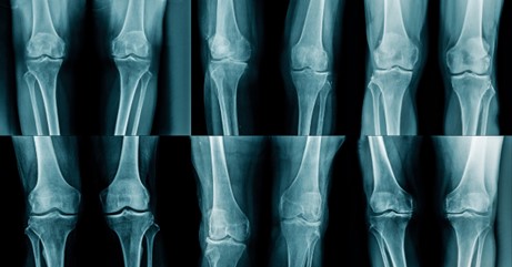
Digital X-Rays have a number of benefits over conventional X-Rays. They produce photos which are clearer and extra detailed, which makes it simpler for docs to diagnose orthopedic situations. In addition they expose sufferers to a decrease dose of radiation, which is a safer possibility. Moreover, digital X-Rays are extra environment friendly as they are often taken and reviewed rapidly, decreasing wait occasions for sufferers.
Along with digital X-Rays, different superior X-Ray applied sciences, corresponding to computed tomography (CT) scans and fluoroscopy, are additionally generally used within the analysis and therapy of orthopedic situations. These superior X-Ray applied sciences present a extra complete and correct understanding of the affected space, permitting orthopedic docs to develop efficient therapy plans and enhance affected person outcomes.
• Magnetic Resonance Imaging (MRI): Magnetic resonance imaging (MRI) is a non-invasive imaging expertise that’s generally used within the analysis and therapy of orthopedic ailments.
MRI makes use of a robust magnetic area and radio waves to supply detailed photos of the bones and joints, which offers a complete view of the affected space. MRI is particularly helpful for visualizing tender tissues, corresponding to muscle tissues, tendons, and ligaments, which can’t be seen on conventional X-Rays. This makes MRI an important device for diagnosing a variety of orthopedic situations, together with accidents to the tendons and ligaments, osteoarthritis, and inflammatory situations corresponding to rheumatoid arthritis.
One of many main benefits of MRI is that it doesn’t use ionizing radiation, making it a safer possibility for sufferers in comparison with different imaging applied sciences corresponding to X-Rays and CT scans. Moreover, MRI permits for the visualization of a number of cross-sectional photos, which may present a complete view of the affected space and assist orthopedic docs decide the perfect plan of action.
Along with diagnostic functions, MRI is used within the therapy of orthopedic situations. MRI can be utilized to watch the progress of remedies and surgical procedures and to trace adjustments within the affected space over time. This info can be utilized to make any essential changes to therapy plans, making certain optimum outcomes for sufferers.
The most recent developments in MRI expertise have improved the standard and velocity of MRI scans. One of the important developments is the usage of high-field MRI machines, which produce higher-quality photos and may detect situations extra precisely. One other development is the usage of quicker MRI machines, which may produce photos in a shorter period of time, decreasing the period of the scan and making the expertise extra comfy for sufferers.
In conclusion, MRI is a invaluable device within the analysis and therapy of orthopedic ailments. Its non-invasive nature, capability to visualise tender tissues and talent to watch the progress of remedies and surgical procedures make it an important device for orthopedic docs. By offering detailed and complete photos of the affected space, MRI performs a vital position in bettering affected person outcomes and making certain the absolute best outcomes for these affected by orthopedic situations.
• Computed Tomography (CT) Scans: Computed Tomography (CT) scans are a sort of medical imaging expertise that’s generally used within the analysis and therapy of orthopedic ailments. CT scans use X-Rays and superior laptop expertise to supply detailed cross-sectional photos of the bones and joints, offering a complete view of the affected space. This makes CT scans an important device for orthopedic docs, as they supply an in depth understanding of the bones, joints, and surrounding tissues, which is essential for the analysis and therapy of orthopedic situations.
CT scans are particularly helpful for diagnosing fractures, dislocations, and different orthopedic accidents that is probably not seen on conventional X-rays. They’re additionally helpful for visualizing the spinal column and sophisticated anatomy, which makes them a invaluable device for the analysis of spinal situations corresponding to herniated discs and spinal stenosis. Moreover, CT scans can be used to information orthopedic procedures, corresponding to biopsies and injections, making certain accuracy and minimizing the danger of problems.
Regardless of the advantages of CT scans, you will need to notice that they do contain publicity to ionizing radiation, which could be dangerous to sufferers over time. Because of this, CT scans needs to be used judiciously and solely when essential. Moreover, the danger of radiation publicity needs to be fastidiously weighed in opposition to the potential advantages of the scan, and various imaging applied sciences, corresponding to MRI, needs to be thought-about when applicable.
The most recent developments in CT expertise have made scans extra environment friendly and correct. One such development is the usage of multi-detector CT machines, which produce photos quicker and with larger element than conventional CT machines. One other development is the usage of low-dose CT scans, which expose sufferers to much less radiation and are a safer possibility.
In conclusion, CT scans are a invaluable device within the analysis and therapy of orthopedic ailments. They supply detailed cross-sectional photos of the bones and joints, permitting orthopedic docs to precisely diagnose and deal with a variety of situations. Whereas you will need to use CT scans judiciously because of the danger of ionizing radiation publicity, they could be a invaluable device for bettering affected person outcomes and making certain the absolute best outcomes for these affected by orthopedic situations.
• Ultrasound: Ultrasound is a non-invasive medical imaging modality that makes use of high-frequency sound waves to supply photos of the bones, joints, and surrounding tissues. This expertise is usually used within the analysis and therapy of orthopedic ailments, because it offers a real-time, dynamic view of the affected space. This makes ultrasound a invaluable device for orthopedic docs, because it offers a transparent understanding of the actions and features of the bones and joints, which is essential for the analysis and therapy of orthopedic situations.
One of many principal benefits of ultrasound is that it doesn’t contain publicity to ionizing radiation, making it a protected and efficient possibility for sufferers of all ages, together with kids and pregnant girls. Moreover, ultrasound is comparatively cheap and could be carried out rapidly, making it a pretty possibility for sufferers who want quick outcomes.
Ultrasound is particularly helpful for the analysis of sentimental tissue accidents, corresponding to sprains and strains, in addition to for the analysis of tendons, ligaments, and muscle tissues. Moreover, ultrasound can be used to information orthopedic procedures, corresponding to injections and biopsies, making certain accuracy and minimizing the danger of problems.
The most recent developments in ultrasound expertise have improved the standard and accuracy of the photographs produced. One such development is the usage of high-frequency ultrasound machines, which produce photos with larger element and accuracy. One other development is the usage of moveable ultrasound machines, which permit docs to carry out scans in quite a lot of settings, together with within the workplace and even on the bedside. This makes ultrasound a handy and accessible possibility for sufferers, particularly those that might have issue touring to a radiology heart.
In conclusion, ultrasound is a invaluable device within the analysis and therapy of orthopedic ailments. It offers real-time photos of the bones, joints, and surrounding tissues, permitting orthopedic docs to precisely diagnose and deal with a variety of situations. Whereas ultrasound does have some limitations, it’s a protected, efficient, and comparatively cheap possibility for these affected by orthopedic situations.
• Bone Scans: Bone scans are a sort of nuclear drugs imaging check that helps to detect adjustments within the bones, corresponding to abnormalities, fractures, infections, and tumors. This check works by injecting a small quantity of radioactive materials into the affected person’s bloodstream, which then accumulates within the bones. A particular digital camera then takes photos of the bones, that are displayed on a pc display.
Bone scans are significantly helpful within the analysis and therapy of orthopedic ailments, as they supply detailed details about the construction and performance of the bones. This info can assist orthopedic docs to establish the reason for ache and different signs, corresponding to swelling and redness, and develop an efficient therapy plan.
One of many principal benefits of bone scans is that they’ll detect adjustments within the bones that is probably not seen on different imaging checks, corresponding to X-Rays or CT scans. This makes bone scans a invaluable device for the analysis of situations corresponding to osteoporosis, bone infections, and bone tumors. Moreover, bone scans can be used to watch the effectiveness of orthopedic remedies, corresponding to bone most cancers remedies and bone fractures, to make sure that the bones are therapeutic correctly.
One other good thing about bone scans is that they’re protected and non-invasive. Not like different imaging checks, corresponding to biopsies or surgical procedure, bone scans don’t contain any incisions or different invasions of the physique. The small quantity of radioactive materials used within the check is eradicated from the physique inside a couple of days, and the publicity to radiation is minimal and regarded protected.
The most recent developments in bone scan expertise have improved the accuracy and effectivity of the checks. One such development is the usage of single photon emission computed tomography (SPECT) scans, which produce extra detailed photos and may detect situations extra precisely. One other development is the usage of hybrid scans, which mix the outcomes of a number of imaging checks to supply a extra complete view of the bones.
In conclusion, bone scans are a invaluable device within the analysis and therapy of orthopedic ailments. They supply detailed details about the bones, which can assist orthopedic docs to establish the reason for ache and different signs and to develop an efficient therapy plan. Whereas bone scans do have some limitations, they’re a protected and non-invasive possibility for these affected by orthopedic situations.
Conclusion:
Orthopedic imaging modalities play a vital position within the analysis and therapy of orthopedic situations. Whether or not it’s X-Rays, MRI, CT scans, ultrasound, or bone scans, every of those imaging modalities has its personal distinctive benefits and can be utilized within the evaluation and administration of orthopedic situations.
to know extra in regards to the rising applied sciences in your trade vertical? Get the most recent market research and insights from BIS Analysis. Join with us at [email protected] to be taught and perceive extra.
[ad_2]
Source link


